Thursday 9 November 2017

VASCULAR ANOMALIES (VA) REGISTRY (PNG)

CREATING A RECORD SYSTEM FOR VASCULAR ANOMALY CASES IN PNG

We appeal to all health workers in PNG to download our single page form below, fill out & then scan & email back to us (molwillie@yahoo.com), with some good photographs of the lesion, for all suspected Vascular Anomalies cases.

Vascular Anomalies are any congenital vascular lesions & a few acquired lesions, composing of either capillaries, veins, lymph-vessels or arteries or a combination of them with or without adjacent tissue overgrowths or hypoplasias. They may be a part of a syndrome or isolated.

For the layperson, if your child is born with an abnormal skin discoloration or swelling, please take them to the nearest Health Care Worker, so that they will be registered & then referred to us for further management.

To Register the case please download the form below, fill & return to us. Contact us for further details & enquiries:

Email: molwillie@yahoo.com PH: 757 16 221 or 715 436 76


We are currently working on our paper to educate all Health Workers in PNG on Vascular Anomalies - Diagnosis & Management. The paper will be posted here, once it is published.

Friday 14 April 2017

Sclerotherapy for Prostate Enlargements

New Break Through Treatment Method for Prostatic Enlargements – Benign Prostatic Hypertrophy (BPH) & Prostate Cancer in PNG: The Common Causes of Urine Outflow Obstruction in Senior Man in PNG

'Sclerotherapy for BPH & Prostate Cancer'

What is Prostate Gland
A Gland located at the urinary bladder outflow that secretes fluids that mixes with & sustains the sperms


What is Prostate Enlargement?

Prostate Enlargements can be caused by 

What are the Usual Treatment Methods?

1. BPHProstate Removal Surgery (Various Procedures/Techniques)
2. Prostate Cancer – Hormonal Treatments or Castration(Bilateral Orchidectomy or Permanent Removal of the Testicles)


Features of Our New Breakthrough Method:


*Achieving Normal Urine Flow & Urination Within 2 Weeks After Just One (1) Single Injection (One Sclerotherapy Session)

*Not Even a Pinch of Blood Lost in this Procedure (Closed Sclerotherapy Method)

*Minimal Pain & Swelling for 3-5 Days (Normal Inflammatory Responses to the Sclerosant)

*Maximum Follow-Up Period (Without Problems & Recurrences) is 5 Years & the Minimum Follow-Up Period, is 4 Months (The Underlying Root Cause of Prostate Enlargement is Dealt With Once & For All)

*Zero (0) Recurrence Rate Among 5 Patients Treated So Far, Currently Alive & Well
-Urinating Normally Without Problems
-Without Intra-Dwelling Catheter (IDC) Re-Insertions
-Without Supra-Pubic Catheters &
-No Hematurias (Bloody Urine)
-No Impotence & Erectile Dysfunctions (No Problems With Libido & Sex)


*Testes Are Preserved; Meaning Fertility, Libido, Erectile & Sexual Reproductive Functions, Remains Much the Same

*Done as an Out-Patient Procedure Meaning, There is No Need for Admission to the Hospital Ward or Can Go Home on the Same Day

*Our Method is a 5 Minutes Procedure!

(All Other Organs & Anatomical Structures Within The Vicinity Remains Preserved or Are Unaltered or Undisturbed)


Disadvantages of Current Standard Treatment Methods – Methods that Deal Directly With the Prostate Gland (Various Prostate Gland Removal Techniques):

-Expensive (Too Much Materials, Technology & Manpower Used for a Poor Outcome Surgery, Especially When Early Transformation of BPH to Prostate Cancer is Missed)
-Many Complications of Prostate Removal Surgery; May Be Life Threatening From Uncontrollable Hemorrhages
-High Recurrence Rates (Narrow Field of View or Sometimes Done Blindly Using a Finger, So Small Microscopic Remains/Spread May Remain & Regrow)
-Long Anesthetic, Operation & Ward Admission Time
-High Risk of Uncontrollable Bleeding
-Numerous blood transfusions may increase risk of blood borne diseases (HIV, Hepatitis B, etc.)
-Recurrence, May Cause a Lot of Problems, Poor Quality of Life for Patients & the High Expenses of Ongoing Incomplete/Unsatisfactory Treatments are Unacceptable in PNG
-In PNG, it is Difficult to Confirm the Early Stages of Transformation to Prostate Cancer (Micoscopic Level Occurrence and/or Spread)

Our Method:

Background & Rational:

It has been known that abnormal back-flow of venous blood carrying a high level of Free Testosterone from the testis, ending up in the Prostate Gland; instead of going out into the systemic flow, is the root cause of Benign Prostate Hypertrophy (BPH) & Prostate Cancer (Y. Gat, et. al.). The amount of Free Testosterone that reaches the Prostate Gland through this abnormal flow is around 130 fold above the systemic level (VERY SIGNIFICANT). The Prostate Gland being constantly exposed to this extremely high levels of Free Testosterone causes it to undergo Hyperplasia (increased number of cells by cell division) & eventually, Transformation into Prostate Cancer. The dependence on hormones to grow is very typical of any Adenocarcinomas (Cancer of Cells that Produce Secretions or Cancers of Glandular Origin). The Prostate Gland not only undergoes Hyperplasia but it is also is full of fluids because of the abnormal venous back-flow. Therefore, there is Edema (tissue swelling because of abnormal amount of fluid accumulation) & a certain degree of Hypertrophy (enlargement/overgrowth of 1 single cell). You can now understand why the Prostate Gland swells up so much to disturb & obstruct the flow of urine.
In Summary, the Root Cause of Prostate Enlargement, is a Varicose Vein of the Internal Spermatic Vein, from Prolonged Upright/Erect Posture, causing back-flow of venous blood, carrying Extremely High Levels of Free Testosterone, flowing Abnormally back into the Prostate Gland. Therefore, we try to deal directly with the venous connection (Deferential Vein) between the Prostate Gland and the Varicose Vein (Internal Spermatic Vein).

With standard 'Prostate Removal Surgical Techniques & procedures', the amount of Free Testosterone within that area is still present (Not Dealt with). If there is a tiny bit of Prostate Gland tissue left behind, the Free Testosterone will stimulate it to regrow abruptly. This is the cause of recurrence with 'Standard Prostate Removal Surgeries', especially when it has already transformed into Prostate Cancer.

Our Technique:
We inject materials (Sclerosants) to cut/block off the main vein (Sclerotherapy) that carries a High Level of Free Testosterone from the Testes to the Prostate Gland that causes/stimulates it to Enlarge & Block the Urine Flow. We achieve this, while preserving the Testes.

We regard BPH & Prostate Cancer as different stages of the same disease; therefore the treatment approach is the same, for both (‘shooting 2 birds with 1 stone’). Both diseases also respond very well to the Removal of the Source of the High Level of Free Testosterone.

*FOR THOSE WITH RECURRENT LATE STATGE PROSTATE CANCER AFTER STANDARD PROSTATE REMOVAL PROCEDURES:

WE ALSO ADMINISTER NEW HORMONAL TREATMENTS FOR SUCH RECURRENT PROSTATE CANCERS, TO SUPPRESS ITS GROWTH & PROGRESSION


THE PATIENT IS HAPPY THAT THE PERMANENT SUPRA-PUBIC CATHETER HAS BEEN REMOVED & HE CAN URINATE NORMALLY AGAIN.

BEFORE THAT HE WAS ONLY WEARING ‘LAPLAP’ NOW HE CAN WEAR A PAIR OF TROUSERS


Call or Email Us Now, For Appointments!

Ph: (675) 715 436 76 or 757 16 221 Email: molwillie@yahoo.com


Tuesday 28 February 2017

Skin Closure/Suture Techniques (Fine Art)


Scarless Suturing


Scarless Skin Closure Techniques

Melanesians have a High Risk for Hypertrophic Scars and Keloids. Therefore, to achieve a scarless closure of any Open Surgical Wounds, carried out on the Melanesian Skin is a great challenge. Most times, we choose Minimally Invasive Techniques such as ‘Sclerotherapy. However, sometimes, it is absolutely necessary to do an Open Surgical Excision to remove any lumps or bumps beneath the skin & then manually suture the opened skin edges together.

Requirements & Guidelines to Achieve Scarless Skin Closure in the Melanesian:

1. Clean/Straight Incisions/Cuts
2. Hemostasis (Stopping of Bleeding) Without Much Use of Diathermy Heat or Use a Bipolar Instead
3. Choice & Size of Suture Materials
4. Choice of Suturing/Closure Technique
5. Anatomic Location of Wound (Tension, Sebaceous Glands, Skin Thickness, etc.)
6. Skin Undermining (tunneling) & Excision of Sebaceous Glands (skin glands that produce oil/sebum)
7. Contour & Skin Color (Avoid ‘Dog Ear’, etc.)
8. Consider Closing Corners First, Inside/Median Side First then Outside/Lateral Side
9. Consider Scar Contractures on Joints (Z-Plasties)
10. Good Diet & Avoid Strenuous Physical Activities, Post Operative
11. Wound Dressing Methods & Timing
12. Avoid Stitch/Suture Marks (Timing of Stitch Removal, etc.)
13. Infection Control (Choice of Antiseptics & Antibiotics)
14. Non-Smoker (Avoid Active & Passive Smoking)
15. Monitoring/Follow-up of Scar & Continued Treatments (Pressure Therapy, etc.)

Notes on Multiple Flaps/Corners for Closure Including Multiple W-Plasties:
1. It takes a lot of time plus effort & may be difficult to incise (execute precisely)
2. Small & Multiple corners on flat surfaces are difficult to stitch-up, have poor blood supply & there is high risk for tip necrosis3. We prefer a straight line closure, blending into natural creases & wrinkle lines & simple & single Z-plasties across/over joint surfaces/creases (perpendicular to)

A Case of a Nose Lump (Exostosis/Osteoma or Benign Bony Outgrowths of the Nasal Bone), Excised & the Skin Edges Were Sutured Using our Finest Suturing Technique. Note the skin darkening from excess skin but a tiny straight scar line.


Another case of Naso-Labial Flap, Donor Site Straight Line Closure Technique.
-The flap is attached to the defect & delayed (left attached) for 3-4 weeks.
-This will allow blood vessels to grow in through the contact skin edges
-After that the flap can be safely divided & the part of the skin covering the defect can survive


The blood supply of the Naso-Labial Flap is from the Facial Artery. During the time of harvest, this artery must be included  within the flap, otherwise there will be flap necrosis (skin becomes black due to no blood supply).


Achieving Almost a Scarless Healing with a Z-Incision for Open Rhinoplasty. A 'Z' Makes it Easy to Incise and to Close Later on, with Good Healing.


We also apply our technique of scarless skin closure to large defects. See here, our technique of closing a scalp (head) defect and also the harvesting of a Latissimus Dorsi Flap to Reconstruct a Neck Defect & the closure of the donor site defect. We prefer a straight line closure.

Scalp Alopecia (Defect) Using a Single Straight Line Closure.


Estimating the tension of skin closure.


Design (Drawing over scar to be excised) & trimming of surrounding hair.


The defect after excision of scar.


Undermining, to reduce tension of skin closure.


Skin edge approximation, to check if undermining is enough (reduced tension).


The defect is closed using a one-layered Nylon, running/continuous  suturing, locked (a knot is tied) at 4 intervals only.


One week after removal of stitches. Take note of the tiny linear scar.


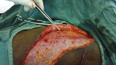
The Harvesting of a Latissimus Dorsi Flap to Cover Defect of the Neck, followed by the closure of a very large donor site defect, using our fine technique. Marking out of the maximum size of skin that can be harvested.


Skin undermining to release/minimize tension.


A very large donor site defect, after complete flap elevation, that needs to be closed.


Partial Donor Site Closure before application of flap, to reconstruct a neck defect.


Appearance of donor site scar after 1 week.


This is the First Case of Latissimus Dorsi (LD) Flap Reconstruction Done in PNG by a Local Surgeon, Using a High Level of Scarless Skin Closure Technique. 

Despite that, there was No Water Running in the Operation Theatre Taps for Scrubbing (Washing Hands). Rain Water Fetched in a Plastic Container the Previous Day & Ordinary Laundry Bar Soap, was provided instead, for Scrubbing (Washing Hands). Despite that, the Surgery Went Well and Its Post-Operative Outcome was Superb.


Before Reconstruction of the Neck Defect (Scar Contracture, which was excised totally) using the above harvested Latissimus Dorsi Flap.


After Reconstruction (covering of the excised scar tissue defect, using the latissimus dorsi flap).


Other Methods for Obscuring Scars:


1. Blending Incision/Suture Lines into Natural Crease & Wrinkle Lines


2. Putting Incision/Suture Lines in Non-Visible Areas
Hiding the Scar Within the Naso-labial Natural Crease Line. 
This patient underwent Open Intra-Arterial Sclerotherapy for Artherio-Venous Malformation of the Upper Lip. The incision is done to visually locate the main artery (feeder) that feeds blood into the swelling, rather than through a catheter; The Superior Labial Branch of the Facial Artery! The Sclerosants convert the vascular malformation into a lump of scar, which was later excise, through the inner part of the upper lip (obscuring/hiding the scar).


It was found by Taylor, a famous Australian Plastic Surgeon, that somehow Arterio-Venous Malformations form along Choke Zones. Choke Zones are borders of Angiosomes (1 area supplied by a single vessel or arterial branch)


Take note that the scars are successfully hidden along the naso-labial crease and the intra-oral side of the lip. Note almost the scarless final appearance of the incision sites as well as the total shrinkage of the Arterio-Venous Malformation.


The Anatomy of the Facial Artery & its Main Branches.

Arterio-Venous Malformation Explained by Dr. William Mol (Vascular Anomalies Specialist).


*Truncal means more central, large vessel (Artery, Vein or Lymph Vessel)

In our patient above, we noted the absence of the superior labial Arterial Branch on the right side only. We then deduced that the branches from the Superior Labial Artery on the Left side connected directly to the branches of the Superior Labial Vein from the Left Side, Without any Capillary Interposition.

Our techniques of scarless surgery, described here, is scheduled to be published in the New PIH e-Med Journal in November 2017.


Most Viewed Postings: